

Studying the structure, integrity, and spatial organization of tissues and cells is one of the keys to understanding the life sciences. In this sense, histology plays a crucial role in biomedical research, because it studies the morphological and biochemical changes that occur in cells during pathological processes.
The aim of histology is to observe and to analyze what is seen under a microscope. In any histological study we proceed in 4 steps :
1) The choise of sample to be studied
2) The techniques to visualize structures and phenomena of interest
3) The production of high quality images
4) The description and interpretation of these images
In order to carry out your histology, imaging and image analysis projects, the platform provides the material, technical and human resources necessary to carry out these projects. Our platform is opened to PARCC teams but also to external, institutional or private teams.
The PARCC histology platform is part of the Histology, Immunostaining and Tissue Imaging (PH2I) platform at Paris Cité University, which also includes the HistIM platform at Cochin Institute (https://www.institutcochin.fr/les-plateformes/histim). Our multisite platform offers future users a complete expertise and a wide range of technologies.
The PARCC histology platform ensures :

Corinne Lesaffre, Facility Manager
Training
The platform has the will to train users so that they can conduct their own studies independently, while benefiting from the experience and technology of this platform. The trained person can then access the different workstations of the platform.
Consulting
The platform can also bring you help in developing your protocols in immunohistochemistry or in improving your different stainings.
Benefit service
The platform can handle your samples at all stages of the histological process according to your request. From paraffin impregnation of tissues through microtome sectioning, staining and immunostaining (brightfield or fluorescence, single or multiple staining) to slide acquisition for quantification and analysis.
Automatic vacuum dehydration (Shandon Excelsior)
Paraffin embedding station (Leica)
2 paraffin microtomes (Leica RM 2145)
3 Cryostats (2 Leica CM 3050 and 1 NX70 MMFrance)
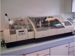
Integrated Workstation (Leica ST2050-CV5030)
Allows standardisation of common stains such as HE – HES – Sirius Red – Masson’s Trichrome – PAS – BA – Orcein.
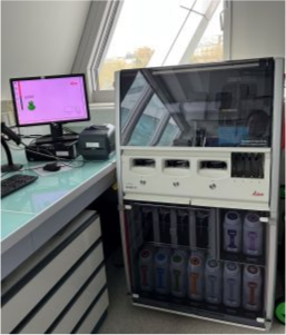
Imunohistochemistry machine (Bond RX Leica)
Fully automated research stainer for ISH, IHC, FISH, RNAscope and Multiplexing in chromogenic and fluorescence. The Bond RX stainer provides the insight for complete control and visibility of each assay. So you can view reagent application, monitor protocol progress, receive comprehensive reports.
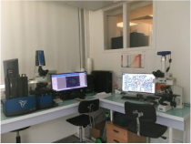
Vectra™and inForm® sofware (Akoya)
Vectra™ is a the system capable of fluorescence multispectral imaging that allow separate up to seven fluorophores from one another and from autofluorescence without cross-talk.
The Vectra system incorporates Akoya’s powerful image analysis software inForm® Tissue Finder and proprietary autofluorescence removal methodology.
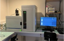
High speed slide scanner for research (VS200 Olympus)
Observation methods: brightfield, fluorescence (DAPI – FITC – CY3 – Texas Red – CY5 – CY7) and Polarisation with Sirius Red for collagen.
Objectives: x20 (NA 0.8), X40 (NA 0.95 and x60 silicon oil immersion (NA 1.42)
Cameras: sCMOS colour camera for brightfield and High sensitivity monochrome camera for the fluorescence
Slide capacity: 210 via 35 trays of 6 slides
Access to the platform services is subject to conditions and management process (according to ISO 9001 standard):
Return and invoicing of services: Once the service has been completed, the applicants are informed by e-mail. The applicant validates the service and signs the follow-up form confirming the end of the service. All services, use of the equipment and training services are subject to a fee and cover maintenance and operating costs. This billing is done every six months. A financial report and a statement of work by team is sent to the platform manager and to the team managers. The equipment is invoiced according to the time reserved. Training and technical services are invoiced according to the techniques requested. The detailed rate for the different services is given at the first meeting. The team leader undertakes to pay the amounts due for the work carried out by the platform.

2017 Imunohistochemistry machine (Bond RX) and 2020 High speed slide scanner (VS200 Olympus)
2021 HALO The image analysis platform for quantitative tissue analysis in digital pathology